Crainio non-invasive intracranial pressure monitor for traumatic brain injury: feasibility study
Crainio Limited · Crainio Limited, Barts Health NHS Trust – Royal London Hospital · Crainio Limited, Research Centre for Biomedical Engineering, City and St George’s University of London
On this page
Abstract
Traumatic brain injury (TBI) affects approximately 70 million individuals annually worldwide, often leading to significant morbidity and mortality. Intracranial pressure (ICP) monitoring plays a vital role in managing severe TBI and other conditions associated with intracranial hypertension by guiding timely medical interventions. However, invasive ICP monitoring requires drilling the skull to place a sensor transducer inside the head, also called a probe. Noninvasive ICP monitoring offers the potential to monitor patients during the critical "golden hour," when timely interventions can significantly influence outcomes. Additionally, it enables continuous surveillance for patients who are not candidates for invasive bolt placement. However, a reliable, continuous, noninvasive ICP measurement remains aspirational despite extensive research efforts. Crainio has developed a noninvasive ICP monitoring system utilising an optical sensor adhered to the forehead. This monitor collects cerebral photoplethysmography signals to estimate ICP changes and detect hypertension, enabling early management and improved patient outcomes. This feasibility study aims to improve the diagnostic precision of Crainio’s algorithm by augmenting the existing dataset, which currently comprises data from 40 TBI patients. A total of 54 participants will be enrolled over one year. Data from simultaneous recordings from the optical sensor and invasive ICP monitors create a more robust dataset to refine and optimise the algorithm for greater clinical accuracy. Additionally, the study will investigate the influence of potential confounders. The ultimate objective is to establish a robust noninvasive monitoring tool to optimize the management of TBI patients, advancing both safety and efficacy in ICP monitoring.
Keywords
- Non-invasive monitoring
- Traumatic brain injury
- Photoplethysmography
- Intracranial pressure monitoring
- Crainio
Introduction
Traumatic Brain Injury (TBI) is the most common cause of death and disability in adults (1). The NHS Emergency Departments admit 700,000 people with head injury per year (2), and roughly 2.5% are severe TBI (3). Current guidelines recommend patients with severe TBI and abnormal CT scans to have intracranial pressure (ICP) monitoring for ~72 hours after injury (4). This is because TBI commonly causes bleeding/brain swelling and, within the confines of the rigid skull, raises the pressure within the skull. High ICP can cause secondary brain injury, increasing disability or causing death (4). Patients for whom guidelines recommend ICP monitoring are approximately 50% more likely to develop elevated ICP due to their condition (5). The full ICP monitoring process costs the NHS ~£9,050/patient (NHS 2022 tariff). In the highest-risk population, this cost is offset by the ability to identify when remedial interventions such as medication, hyperventilation, cooling, craniotomy or draining cerebrospinal fluid, among others, are required to prevent further damage to the brain. In the UK, ~16% of patients with severe TBI (~2,800/annum) receive ICP monitoring (6)(10). This may be ~43% of the patients that guidelines recommend (1). This low compliance may be explained by the fact that existing methods are extremely invasive, resource-intensive, and logistically challenging: a neurosurgeon must insert an electrical sensor directly into brain tissue through a small hole drilled in the skull. Complications can occur and the crucial “golden hour” for monitoring is missed (7), delaying treatment and therefore increasing the risk of poorly outcomes. Other patients have their ICP “monitored” using frequent CT/MRI scans, the sensitivity of this qualitative method for detecting raised ICP is widely acknowledged to be low (7,8). The usefulness of this method is also low because the imaging can only capture a snapshot view of the brain instead of a continuous view, and imaging waiting times mean the “golden hour” is still usually missed. Logistical challenges, costs and risks of invasive ICP monitoring leave>3,700 of the highest-risk patients/annum with no or substandard monitoring, while continuous surveillance for patients who are not candidates for invasive bolt placement is not even an option in the current clinical management pathway. Crainio is developing a ground-breaking non-invasive ICP (nICP) monitor comprising an optical sensor stuck to the forehead, a control unit and a monitor. The transformative technology shines harmless near-infrared light (NIR) through the skull and into the vessels on the surface of the brain to detect a reflected optical pulsatile signal known as photoplethysmography (cerebral PPG). When the ICP is high (ICH), the transmural pressure constricts the vessels on the surface of the brain at low vascular tone sites, leading to morphological changes in the cerebral PPG signals. These are analyzed through quantifiable morphological features, enabling the development of a computational model that can estimate high ICP non-invasively.
Novel non-invasive methods for continuous ICP monitoring have begun to be implemented in clinical practice, including skull deformation waveform analysis, ultrasound-based techniques, and tympanic membrane pressure measurements. These emerging modalities highlight both the clinical demand and the growing recognition of the need for reliable non-invasive systems to support the early detection and treatment of intracranial hypertension. Within this context, cerebral photoplethysmography (PPG) represents a promising approach, offering a direct physiological signal from cerebral microcirculation and the potential to overcome limitations associated with some existing alternatives (9,10). Crainio's small, portable and non-invasive form could extend beyond intensive care units, providing critical information to healthcare practitioners in pre-hospital or emergency department settings. Also, it could massively increasing the number of TBI patients who receive continuous ICH monitoring, enabling earlier monitoring and thus earlier and better informed intervention, improving patient outcomes. Crainio’s scientific background has shown encouraging results in an adult head phantom (11–14), adult healthy volunteers and 40 adult patients with severe TBI in ICU (15–19), are proof that cerebral PPG feature-based algorithms can estimate ICP in a population with TBI. Nonetheless, as a ML-based technology, the extension of the PPG-based ICP monitoring dataset will enhance the model’s accuracy on a diverse population where possible optical confounders are evaluated (i.e. skin pigmentation, skull density, skull thickness). To generate a strong database for enhancing the Crainio model’s accuracy, this protocol paper describes a feasibility study for collecting simultaneous non-invasive and invasive signals from severe TBI patients. Researchers will analyse the effect of possible confounders for light-tissue interactions, such as skin pigmentation, skull thickness and age. This protocol paper followed the Standard Protocol Items: Recommendations for Interventional Trials (SPIRIT) checklist (Appendix) to report relevant clinical studies details as recommended by the EQUATOR Network (Enhancing the Quality and Transparency of Health Research). Purpose and aims Purpose: Collect cerebral photoplethysmogram signals and concurrent invasive ICP measurements from patients with traumatic brain injury to develop Crainio machine learning algorithms to the point that they can detect raised intracranial pressure (ICP>20 mmHg) with sufficient sensitivity and specificity that Crainio monitor can be considered for clinical use. ● Aim 1: Collect the algorithm training and testing data with which to improve the accuracy of the Crainio non-invasive intracranial pressure monitor to levels that will be acceptable for use in clinical care. ● Aim 2.1: Determine any undesirable side effects of the Crainio monitor. ● Aim 2.2: Identification of the effect of confounders in Crainio measurements accuracy. ● Aim 2.3: Assess the impact of patient and usability factors on the monitor’s performance.
Methods
Study design This is a single-arm study in which both optical and invasive monitoring are conducted simultaneously for all participants. As a result, intervention randomization and blinding are not required, as all participants receive the same standardized procedure for data collection. However, during the off-line analysis of the optical signals, data will be randomly assigned to the training or the testing group. This study has been approved by the Medicines & Healthcare Products Regulatory Agency (MHRA) reference CI/2024/0045/GB and the HRA and Health and Care Research Wales (HCRW), REC reference 24/LO/0554. Moreover, this study is prospectively registered on clinicaltrials.gov (NCT03594734). The protocol was designed following the ISO 14155 standards for good clinical practice, and the research team has been certified in GCP. The final protocol, version 6.0, was updated in October 2024, and amendments were notified to the regulatory entities. Study setting All study procedures will take place at the Royal London Hospital (RLH), Barts Health NHS Trust, United Kingdom. Participants and recruitment Participants will be identified when they are admitted to the Royal London Hospital and require invasive ICP monitoring. There are between 100-150 suitable patients admitted each year at the Royal London Hospital. The neurosurgical registrar on call will screen any potential participant, including against the sample size requirements based on age and skin colour. Recruitment will take place over a 12-month time period to ensure the target sample size is reached. Once a potential participant is identified, if unconscious, the relatives will be approached by the registrar to inform them of the study. If relatives are not available, a professional consultee not involved in the study will be approached and asked for assent for the patient’s participation. A declaration of no objection for the patient to participate in the study will be sought from these individuals. In the minority of cases where the patient is conscious, he/she will be approached by the clinician directly.
Figure 1. Study flow diagram. 5
| Inclusion Criteria | Rationale |
|---|---|
| 16–99 years of age | Young people and adults range as defined by the NHS. The bone density of the skull varies from young people to older adults, which is one of the confounders to investigate in this research. |
| TBI diagnosis | TBI is the primary cause of intracranial hypertension worldwide, enabling the collection of data at normal and high ICP. |
| Invasive ICP monitoring | Patients who are having invasive ICP monitoring as part of their normal medical treatment. The reference for training Crainio's algorithm is the invasive ICP measurement. |
| Exclusion Criteria | Rationale |
| The forehead skin is not intact | The probe is stuck in the forehead using a medical adhesive. Skin integrity is required to place the sensor. |
| Decompressive craniectomy | Due to the science behind the monitor, the skull must be closed. |
| Open external ventricular drainage | When the EVD is open, there are no valid recordings of invasive ICP. |
| Unlikely to survive the following twelve hours | The duration of the study is 12 hours, corresponding to the time when the acquisition of both the invasive ICP measurements and the cerebral PPG signal collection is started. |
| Known acrylates allergy | The probe's adhesive contains acrylates, which might lead to a skin rash in patients allergic to this material. |
Eligibility
The principal investigator (PI) will determine the patient's eligibility. Eligibility criteria and rationale are summarized in Table 1. Informed consent will be obtained by the PI in a private setting at RLH prior to any research procedures taking place.
Recruitment will occur in a high-acuity hospital environment, with procedures designed to ensure ethical compliance and feasibility. In accordance with the Declaration of Helsinki, unconscious patients may be enrolled without prior consent. Relatives will be approached at an appropriate time to provide assent; if relatives are unavailable or assent is inappropriate, a professional consultee (a treating physician not involved in the study) will be consulted. Patients who regain capacity during the monitoring window will be informed of their participation and invited to provide deferred consent, while those who remain without capacity until discharge will not be informed. Conscious patients undergoing invasive ICP monitoring will be approached upon hospital admission, with interpreters available if required. Consent will be obtained by the neurosurgical registrar, and participants will have up to 24 hours to decide. Enrollment will begin only once written consent or assent has been obtained. Data collection will be performed by the research team identified in the ethics protocol, with recruitment overseen by the steering committee. The research team has prior experience implementing these procedures, having successfully conducted a pilot study involving 40 patients with traumatic brain injury, which demonstrated the feasibility of the recruitment and consent strategy (7–9).
Participants may decide to withdraw themselves from the investigation. This is possible for participants who are conscious during their participation in the study or wake up during the monitoring hours. Acquisition of study data will stop as soon as they withdraw consent.
Regardless of the circumstances of withdrawal from the study, all study data collected about a withdrawn participant will be removed from the study if the participant asks for it, but retained in accordance with research and trust data governance.
Intervention
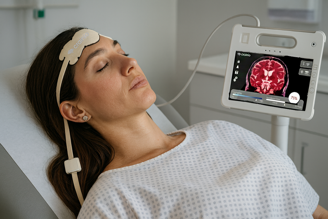
Reference measurement After obtaining consent or assent, the patient will be enrolled in the study. The registrar or consultant on call will be responsible for the insertion of the invasive ICP sensor as a standard procedure in these cases. In the RLH, a parenchymal ICP system (Neurovent-p, RAUMEDIC AG) is used to monitor TBI patients who receive an invasive ICP probe, which is connected to a BeneVision N1 module for Mindray monitors (MINDRAY BIO-MEDICAL ELECTRONICS CO., LTD, China). All relevant waveforms from the multifunctional monitor connected to the patients will be recorded with the ICM+ software (ICM+, University of Cambridge, UK) on an exclusive research laptop allocated to this study. Crainio acquisition monitor The end-to-end Crainio monitor that features in this study is composed of three main components: the disposable sensor, the control unit (instrumentation), and software (nICP monitor) running on a standard third-party laptop device in tablet mode (Figure 2). The sensor is positioned on the patient’s forehead and is used to shine light into the brain and detect the light coming back from the brain. The sensor is connected to the main control unit at the opposite end. The sensor represents the front-end of the monitor and it will be in contact with the patient’s forehead for the duration of the measurement. The primary function of the sensor is to shine near-infrared light into the patient’s brain. This is achieved by three light emitting diodes (LEDs) emitting light in the near-infrared range of the spectrum, one at a time. Figure 2. Crainio acquisition monitor.
The light scattered back from the brain is detected by two photodiodes with large active areas to maximise the sensitivity. Light diffuses in tissues in an arc trajectory from the light source to the detector (20), with the depth of penetration proportional to the separation distance between the source and detector. The photodiodes are placed at different distances from the LEDs package. The proximal photodiode in the nICP sensor is placed at 10 mm from the LEDs and detects the light interrogating shallow tissues such as the skin, skull and extracerebral tissues. The distal photodiode is positioned at 30 mm from the LEDs and detects the light interrogating a deeper tissue volume like the brain surface. The control unit is responsible for driving the light sources (LEDs), amplifying the signals detected by the photodiode, removing the ambient light contamination, pre-processing the signals and digitizing them for use by the nICP monitor. It is a small, rugged unit that connects to the sensor via a flexible cable, and to the laptop via USB cable. The nICP monitor displays the signals and saves the measurements in a readable text file. These measurements will be used for further development of the Crainio machine learning algorithms, as per the Primary Objective of this study to continue to improve the accuracy with which this monitor can detect intracranial hypertension. Clinical procedure Patients who fit the inclusion criteria will be enrolled in the study by the neurosurgical team looking after These patients will already have invasive ICP monitoring in place or will have this placed as part of their standard treatment. Once invasive ICP monitoring is in place, a member of the clinical team will place a Crainio sensor on the same hemisphere of the forehead (frontal lobe) where the invasive probe is located (Figure 3). Once they have positioned the Crainio sensor using the standard adhesive and a loose head band, they will connect it to the control unit via the flexible cable. Figure 3. Probes placement. Once the connection between the control unit and the computer has been established, acquisition can commence in the nICP monitor. After assessing a satisfactory quality of the
signals, the recording of PPG signals will commence. The recording of these signals will continue for up to 12 hours (while the invasive ICP monitoring is in place). Simultaneously to the collection of cerebral PPG signals, vital signs from the multifunctional monitor connected to the patients will be recorded with the ICM+ software into the laptop. Initial movement artifacts generated by three taps on top of both invasive and non-invasive monitors’ cables will allow off-line signal synchronization. The location of the skin where the Crainio sensor is positioned will be regularly checked by the clinical staff for skin integrity. At the end of the 12 hours period, or if the invasive ICP monitoring is removed, the recording of cerebral PPG signals will be stopped and the Crainio sensor removed and disposed of at the correct biohazard bags. While the Crainio monitor plays a pivotal role in this research, all clinical decisions regarding patient care will continue to rely solely on established clinical ICP measurements. In this study, the Crainio monitor serves as a valuable research tool for data collection and analysis only. Ultimate responsibility for patient care and medical decision-making will remain firmly rooted in the clinical assessment of ICP by healthcare professionals using standard of care.
Results
All patient information and measurements collected in this study are summarized in Table 2. Information will be obtained from the medical record as sex, age, cause of trauma, medication, and Glasgow Coma Scale (GCS). Skin pigmentation will be measured in the forehead using the individual typology angle and melanin index provided by the Colorcatch (Delfin Technologies, Finland). Data from the CT scan regarding oedema severity, ventricles engorgement, skull thickness below the sensor, and presence of haemorrhages will be recorded by the consultant. Additional vital signs such as arterial blood pressure and finger oxygen saturation will also be acquired simultaneously from the Mindray monitor. Finally the parameters from the ventilator will be recorded as well as the results of all blood gas analysis during the nICP monitoring time. The acquisition of the ICP measurements, and cerebral PPG signals from the patient will continue for a total of up to 12 hours, including during those periods where the patient’s position is changed. Depending on the individual circumstance of the patient, this may be continuous or intermittent periods totalling 12 hours. Clinical staff will perform their standard regular checks on the patient as the regular medical treatment. A Co-investigator from Crainio may be present in the intensive care unit at times. They will monitor the data acquisition in terms of signal quality, correct saving of the data and battery duration on an ad hoc basis. The Co-investigator will record the data and will log-events on the electronic clinical output form. The Co-investigator from Crainio will have an honorary contract with Barts Health NHS Trust and up to date GCP training. Once the measurements have been acquired for 12 hours, the acquisition will stop and the Crainio sensor will be removed. The clinical team will evaluate whether the patient requires the continuation of invasive ICP measurements. The performance of the algorithms developed from the collected measurements will be comprehensively assessed using a range of accuracy metrics. These include sensitivity, specificity, positive predictive value, and negative predictive value to evaluate classification capabilities. Additionally, for continuous models, metrics such as mean absolute error (MAE),
| Category | Details |
| Intervention measurements | 1. Cerebral PPG signals (Crainio monitor); 2. Invasive ICP monitoring as part of their medical treatment (ICM+ software for Mindray monitors), waveforms. Device: BeneVision N1 module for Mindray monitors and Neurovent p probes for ICP monitoring; 3. Crainio probe location (left-right) (Observation from PI); 4. Invasive ICP probe location (left-right) (Observation from PI) |
| Demographics | 5. Age (Clinical record); 6. Sex (Clinical record); 7. Cause of trauma (Clinical record); 8. Skin pigmentation/colour (SkinColorCatch, Delfin Technologies) |
| Clinical parameters | Glasgow Coma Scale (GCS) (Clinical record); Medications during recording time (Clinical record); Ventilator parameters (Ventilator as part of their medical treatment). Device reference ServoU, Maquet Getinge; Events such as: aspiration, body position change, medication; Invasive arterial blood pressure (ABP) monitoring as part of their medical treatment (ICM+ software for Mindray monitors), waveforms; Finger blood oxygen saturation (SpO2) monitoring as part of their medical treatment (ICM+ software for Mindray monitors) waveforms |
| Photographs/videos | Photographs/videos taken with the laptop camera and stored in a password-protected folder. These can potentially be published in a manner that ensures the patient's anonymity |
| Laboratory assessments | Blood gas analysis (Arterial blood gas as part of their medical treatment). Device: ABL90+, Radiometer |
| Radiology assessments | Skull thickness (CT scan as part of their medical treatment); AIS severity classification from imaging (CT scan as part of their medical treatment); Helsinki score for grading the severity of the injury (CT scan as part of their medical treatment); Ventricular engorgement by Evan's index (CT scan as part of their medical treatment); Presence of haemorrhage and location (CT scan as part of their medical treatment) |
root mean square error (RMSE), bias, and limits of agreement will be employed. This multifaceted approach ensures robust evaluation across different algorithm types, providing a clear understanding of their clinical reliability and precision. Sample size The sample size was determined based on the study’s primary objective of improving the accuracy of the Crainio non-invasive intracranial pressure monitor to levels that will be acceptable for use in clinical care. Assuming that the true sensitivity and specificity would each be approximately 90%, that the 95% confidence interval around these estimates should be no wider than ±5%, and that the algorithm must demonstrate performance exceeding a clinically relevant minimum threshold of 80% with at least 80% power. Because each participant contributes multiple 60-second data windows, which are correlated within individuals, paired measurements every 12 minutes are used for testing the algorithm, leading to a conservative intraclass correlation of 0.25 was applied to account for within-patient clustering. It was also assumed that approximately 18% of windows would reflect raised ICP [7] and that up to 15% of participants may not complete the planned monitoring period due to clinical or operational factors. A binomial precision criterion was applied to ensure that sensitivity and specificity are estimated with sufficient statistical accuracy, while a superiority test was used to ensure that the algorithm’s performance is demonstrably better than a clinically relevant minimum threshold. Under these assumptions, a total of 54 participants are required to achieve the statistical precision and power targets for both sensitivity and specificity. To allow for a standard 70:30 split between training and testing datasets, the study will recruit 54 participants, with approximately 38 allocated to algorithm training and 16 allocated to independent performance testing. This number is sufficient to meet the study Secondary Endpoints too. Each skin tone group should have at least 20% of the participants. This is because regulators are now requiring medical devices employing PPG technology to be validated on a population where at least 20% of the study participants have dark skin tone (21). Moreover, it would ensure that Crainio algorithms are responsive to all skin pigmentations. Additionally, at least 20% of the study participants (12) should be under 25 years old and at least 20% (12)should be more than 45 years old. This serves to ensure a wide range of skull densities to develop a robust model, as skull density is correlated with age (22).
Table 2. Study flow diagram.
● Glasgow coma scale (GCS) (Clinical record) ● Medications during recording time (Clinical record) ● Ventilator parameters (Ventilator as part of their medical treatment). Device reference ServoU,
Maquet Gettinge
The patient’s routine radiological examinations of the brain will be looked at ● Skull thickness (CT scan as part of their medical treatment) ● AIS severity classification from imaging (CT scan as part of their medical treatment) ● Helsinki score for grading the severity of the injury (CT scan as part of their medical treatment) ● Ventricular engorgement by Evan’s index (CT scan as part of their medical treatment) ● Presence of haemorrhage and location (CT scan as part of their medical treatment)
Allocation
Random allocation on patients’ level partition will take place in the analysis phase, where 70% of the patients (i.e. 38 patients) will be allocated to the training group for fitting the model, from which 20% will be used as a validation set for hyperparameters tuning, model selection and generalization. The reserved data from the remaining 30% of the patients (i.e. 16 patients- kept separate and completely unseen from training/validation phase) will be used to test the model's accuracy in terms of sensitivity, specificity, positive prediction value, and negative predictive value. Data management and quality assurance All study data will be collected and managed following rigorous standards to ensure data security, integrity, and compliance with relevant regulations as shown in Figure 4. PPG signals will be collected using the Crainio probe, controlled by the Control Unit, and transferred via the nICP monitor on a dedicated study laptop to a designated OneDrive folder. Data will be saved directly to the cloud, ensuring no local data resides on the laptop. Invasive ICP and vital signs data will be collected using the hospital’s Mindray monitor, managed by hospital staff, with the ICM+ software transferring the data directly to the same OneDrive folder. Demographics and medical history will be accessed by the principal investigator and
recorded using ePRO forms designed in the EPI Info package.. All data entry will be reviewed at the end of each session to confirm accuracy before being uploaded to the secure cloud folder by the research team. Figure 4. Processing and storage of the study data. In compliance with UK clinical trial regulations, datasets will be archived securely for up to 10 years. Statistical analyses and quarterly reports summarizing data quality and study progress will be prepared and presented to the study steering committee. The steering committee comprises an independent clinician, clinical governance member, and patient and public representative. Statistical methods General signal processing will be performed using Python. PPG signals are known to be sensitive to various noise sources, such as movement. Therefore, movement artifact anomalies and photodetector saturation sections will be removed from the cerebral PPG signals, along with the respective synchronous segments from the invasive ICP reference signals. The cerebral PPG signals will then be filtered using Butterworth filters to separate the AC PPG component from the DC PPG component. The filtered signals will subsequently be normalized by dividing the AC part of the signal by its DC component. Several morphological features that have been investigated in the literature to characterize pulsating signals, such as PPG (23), will be extracted. This study will extract more than 100 time-domain and frequency-domain features from the cerebral PPG signals and their derivatives. The median value of each feature will be calculated over a signal window of 60 seconds. Similarly, the median value of invasive ICP will be calculated for this period. The dataset of
patients will be randomly divided into training and testing sets, particularly, 38 patients will be allocated for the training and validation phase, and the remaining 16 patients will be reserved for the test set to assess model performance on unseen data. Of the training set, 20% will be held out for fine tuning models’ hyperparameters; this group will be referred to as the validation set. Both the training and validation sets will be used to train, configure and select best machine-learning models. All patients from the previous clinical proof-of-concept study will be included in the training group. Features will be ranked and selected to optimize a parsimonious model, this includes removing collinear pairs to reduce redundancy and computational complexity as well as feature elimination using recursive feature elimination with wrapped Random Forest Model Features will then be fed into classification models for estimating normotensive and hypertensive classes, and a later stage regression models will be evaluated as well. Both classical ML models and Neural Networks (NN) will be employed. Classical ML models, namely, Random Forest, AdaBoost, LGBM, SVM and KNN, will be used as baseline models for comparisons. As for NN models, variants of Recurrent Networks (RNN) to Convolutional Networks (CNN) and combinations of the two types will also be developed, such as LSTM/GRU and Temporal Convolutional Network, ResNet, Unet and other variants. Attention layer and residual connections will also be incorporated to capture and enhance extraction of both temporal and spatial domain variations in the data to improve our estimation. The arterial blood pressure waveform, clinical parameters, demographic data, and relevant events will be included in the model analysis. These inputs will offer a comprehensive framework for understanding the relationship between non-invasive intracranial pressure estimates and actual intracranial hypertension. Potential confounders, such as age, skin pigmentation, and variations in skull thickness, will be identified and examined through subgroup analyses. These analyses will evaluate how these factors influence the performance of the nICP model. Bland-Altman analysis will be conducted to determine inter-method agreement, adjusted for the extensive number of repeated measurements per patient. The correlation between methods will be assessed using the Pearson correlation coefficient. Receiver operating characteristic (ROC) curves will be constructed to evaluate the sensitivity, specificity, positive predictive value (PPV), negative predictive value (NPV), and area under the curve (AUC) of the estimated values compared with the measured values for elevated ICP. Statistical significance will be defined as a p-value < 0.05. These statistics will be calculated from pairs of observations (invasive and non-invasive ICP),treating each pair as an independent measurement. For classification models, pairs of observations will be categorized as normotensive or hypotensive.
Harms
The optical sensor will be affixed to the forehead of neurocritical care patients with traumatic brain injury (TBI) for up to 12 hours. During this time, near-infrared light will be emitted into the tissue using a controlled current of up to 50mA. This non-invasive tool is associated with a low risk of unintended consequences or adverse effects. Participants may experience mild, temporary marks on the skin after the sensor is removed. However, no skin lesions, burns, or other lasting effects are anticipated. Any marks that do appear are expected to fade quickly, leaving no permanent traces on any participant.