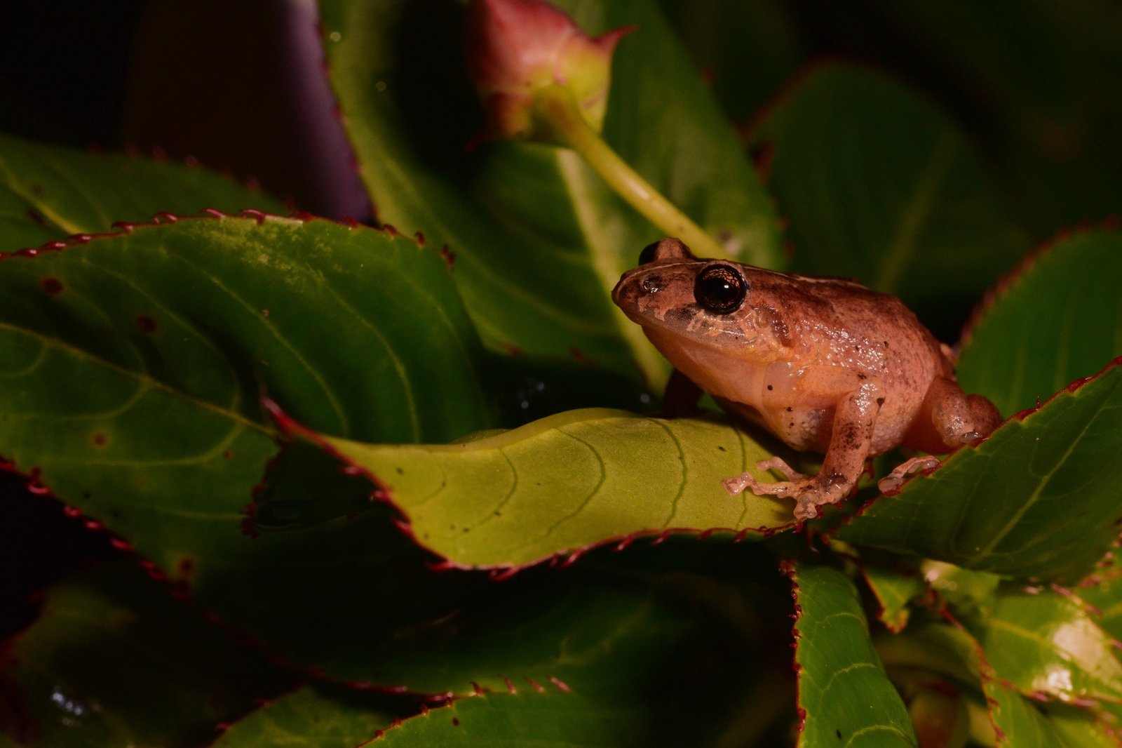
Dorsal design variation and melanin associated pigmentation in preserved specimens of Pristimantis bogotensis (Anura: Craugastoridae)
Universidad Javeriana, Colombia
On this page
Abstract
Phenotypic variation in dorsal design patterns and pigmentation represents an important component of amphibian diversity and may be influenced by genetic, developmental, and environmental factors. However, the characterization of these traits using preserved specimens requires careful interpretation because fixation and long-term storage can alter the appearance of some chromatophore-derived pigments. Pristimantis bogotensis, an endemic frog from the Eastern Cordillera of Colombia, exhibits substantial dorsal variation, with previous observations describing 11 dorsal patterns in living individuals from Chingaza National Natural Park. In this study, we characterized dorsal design variation and preserved pigmentation in 290 specimens of P. bogotensis deposited in the Museo Javeriano de Historia Natural “Lorenzo Uribe, S.J.” (MPUJ). Standardized photographic documentation and digital image analyses were used to identify dorsal design categories, describe preserved pigmentation, and quantify melanin-associated dark pigmentation. A total of 17 dorsal design patterns were recognized, expanding previous descriptions of the species. Dark pigmentation coverage was quantified using image thresholding, and its relationship with body size and elevation was explored. The analyses revealed no significant association between melanin-associated pigmentation and either snout–vent length or elevation. Although preserved pigmentation displayed a wide range of visible tones, these observations should not be interpreted as an exact representation of the coloration of living individuals due to potential alterations associated with preservation methods. Nevertheless, the documented dorsal patterns and melanin-associated pigmentation demonstrate the value of biological collections for investigating morphological diversity when preservation-related limitations are considered This study provides the most comprehensive characterization of dorsal design variation currently available for P. bogotensis and establishes a morphological baseline for future studies integrating live observations, ecological information, and genetic approaches to evaluate the mechanisms underlying phenotypic variation in this species.
Keywords
- Anura
- cryptic coloration
- polychromatism
- perceptual switching
- image threshold
Abstract
Phenotypic variation in dorsal design patterns and pigmentation represents an important component of amphibian diversity and may be influenced by genetic, developmental, and environmental factors. However, the characterization of these traits using preserved specimens requires careful interpretation because fixation and long-term storage can alter the appearance of some chromatophore-derived pigments. Pristimantis bogotensis, an endemic frog from the Eastern Cordillera of Colombia, exhibits substantial dorsal variation, with previous observations describing 11 dorsal patterns in living individuals from Chingaza National Natural Park.
In this study, we characterized dorsal design variation and preserved pigmentation in 290 specimens of P. bogotensis deposited in the Museo Javeriano de Historia Natural “Lorenzo Uribe, S.J.” (MPUJ). Standardized photographic documentation and digital image analyses were used to identify dorsal design categories, describe preserved pigmentation, and quantify melanin-associated dark pigmentation. A total of 17 dorsal design patterns were recognized, expanding previous descriptions of the species. Dark pigmentation coverage was quantified using image thresholding, and its relationship with body size and elevation was explored.
The analyses revealed no significant association between melanin-associated pigmentation and either snout–vent length or elevation. Although preserved pigmentation displayed a wide range of visible tones, these observations should not be interpreted as an exact representation of the coloration of living individuals due to potential alterations associated with preservation methods. Nevertheless, the documented dorsal patterns and melanin-associated pigmentation demonstrate the value of biological collections for investigating morphological diversity when preservation-related limitations are considered.
This study provides the most comprehensive characterization of dorsal design variation currently available for P. bogotensis and establishes a morphological baseline for future studies integrating live observations, ecological information, and genetic approaches to evaluate the mechanisms underlying phenotypic variation in this species.
Introduction
Variation in coloration and dorsal design patterns is a widespread phenomenon among amphibians and represents an important source of phenotypic diversity. In anurans, individuals within the same species may differ in background pigmentation and the presence, arrangement, and intensity of stripes, spots, and other dorsal markings. Such variation may influence ecological interactions, including camouflage, predator avoidance, thermoregulation, and communication (Duellman & Trueb, 1986; Stevens & Cuthill, 2006). However, the adaptive significance and mechanisms responsible for maintaining this diversity remain unresolved for many species (Hoffman & Blouin, 2000; McKinnon & Pierotti, 2010).
The expression of pigmentation and dorsal patterns can be influenced by multiple factors, including genetic variation, developmental processes, and environmental conditions. In amphibians, color and pattern polymorphisms may result from heritable differences, phenotypic variation, or interactions between genotype and environment (Summers et al., 2004; Schlichting & Pigliucci, 1998; Crispo, 2007). Consequently, documenting the extent and distribution of phenotypic variation within natural populations represents an essential first step toward understanding the ecological and evolutionary processes underlying these traits.
Amphibian coloration is generated by specialized pigment cells known as chromatophores, including melanophores, xanthophores, and iridophores, which produce dark melanins, carotenoid- and pteridine-based yellow and red pigments, and structural coloration (Bagnara et al., 1968; Rodríguez et al., 2020). Melanin is one of the principal pigments responsible for dark coloration in amphibians and can play multiple biological roles, including thermoregulation, ultraviolet radiation protection, and visual signaling (Bagnara et al., 1968; Falk et al., 2026). Nevertheless, the study of coloration using preserved specimens requires caution because fixation and long-term storage can alter chromatophore structure and modify the appearance of some pigments (Juszczyk, 1952). Despite these limitations, preserved specimens can retain valuable information related to dorsal design elements and melanin-associated pigmentation, making biological collections useful resources for studying morphological variation across broad temporal and geographic scales.
Pristimantis bogotensis is an endemic Andean frog distributed in the Eastern Cordillera of Colombia, where it inhabits high-Andean forest and páramo ecosystems. Previous observations by Hoyos (1991) documented considerable dorsal variation in living individuals from Chingaza National Natural Park, describing 11 dorsal design patterns. However, a quantitative characterization of dorsal design variation and preserved pigmentation across a larger series of museum specimens has not been conducted.
The present study examines preserved specimens of Pristimantis bogotensis deposited in the Museo Javeriano de Historia Natural “Lorenzo Uribe, S.J.” (MPUJ) to characterize the diversity of dorsal design patterns and melanin-associated pigmentation present in the collection. Additionally, we explore whether the extent of dark pigmentation is associated with body size and elevation. This study provides a morphological baseline for future research integrating live observations, ecological information, and genetic approaches to evaluate the mechanisms underlying phenotypic variation in this species.
Methods
Specimen selection and examination
A total of 334 preserved specimens identified as Pristimantis bogotensis and deposited in the Museo Javeriano de Historia Natural “Lorenzo Uribe, S.J.” (MPUJ), Colombia, were initially examined. Specimen-associated information, including collection locality, geographic coordinates, elevation, and collection date, was obtained from the digital records available through the Global Biodiversity Information Facility (GBIF) database. Species identification was verified through direct examination of each specimen based on external morphological characteristics and comparison with available taxonomic descriptions.
Each specimen was examined to assess its preservation condition, developmental stage, and suitability for subsequent analyses. The developmental stage was classified as juvenile or adult using a combination of available museum information, body size (snout–vent length, SVL), and the presence of external secondary sexual characteristics when discernible. Sex determination was not included in this study because reliable identification of sex in preserved specimens often requires examination of internal reproductive structures, and invasive procedures such as dissection were not performed to preserve the integrity of the biological collection.
Specimens showing severe deterioration, loss of relevant dorsal structures, poor preservation, or conditions preventing reliable identification of dorsal design patterns were excluded from the analyses. After applying these criteria, the final dataset consisted of 290 specimens (Figure 1). The examined specimens had experienced different preservation histories, including formalin fixation followed by transfer to ethanol and direct preservation in ethanol. Because preservation methods can differentially affect the appearance of chromatophore-derived pigmentation, the analyses were interpreted as descriptions of preserved pigmentation rather than the original coloration of live individuals.
Image acquisition
All specimens were photographed under standardized laboratory conditions using a Nikon D5200 digital camera (24 megapixels; maximum resolution 6000 × 4000 pixels) attached to a Nikon SMZ1270 stereoscope. During imaging, specimens were placed individually in Petri dishes containing 70% ethanol to minimize surface reflections and maintain a consistent orientation. To prevent movement or floating of specimens during photography, individuals were gently stabilized using a fine needle supported by modeling clay positioned on the rim of the Petri dish. Images were obtained using a standardized setup consisting of a blue background, two white LED light sources positioned laterally, and a halogen light source placed behind the Petri dish to reduce variation in illumination among photographs. Dorsal and ventral images were obtained for each specimen. For larger individuals that exceeded the stereoscope field of view, multiple images were taken to document the complete ventral surface. An additional image of each specimen was taken over graph paper to provide a scale reference for morphometric measurements.
Figure 1. Specimen selection process from the original sample.
Morphometric measurements and developmental assessment
Snout–vent length (SVL) was measured from digital images using the straight-line measurement tool in ImageJ software. The scale was calibrated using a known distance of 10 mm from the graph paper image. Measurements were taken from the tip of the snout to the posterior margin of the cloacal opening.
SVL measurements were used as one of the criteria supporting developmental stage classification in conjunction with museum records and external secondary sexual characteristics when visible. Because sexual dimorphism may influence body size and external morphology in amphibians, the absence of sex determination represents a limitation that should be considered when interpreting size-related analyses.
Dorsal design pattern characterization
Dorsal design patterns were characterized through visual examination of standardized dorsal photographs. The classification was based on the presence, arrangement, and relative distribution of visible dorsal markings, including stripes, bands, spots, and other contrasting pattern elements. The resulting pattern categories represent descriptive morphological classifications based on preserved specimens and should not be interpreted as discrete genetic morphs. Individuals for which dorsal markings could not be reliably distinguished due to preservation artifacts or specimen deterioration were excluded from the analysis.
Characterization of preserved pigmentation
The preserved pigmentation visible on dorsal photographs was documented using Adobe Color (2021) to obtain standardized digital color palettes. Five points were selected across the dorsal surface of each specimen, and the corresponding color values were recorded to describe the range of pigmentation retained after preservation. Because preservation methods, including formalin fixation and long-term storage in ethanol, can alter the appearance of some chromatophore-derived colors, these data were interpreted as a representation of preserved pigmentation rather than the complete coloration of living individuals. The extracted digital color values were compared with the color catalogue for field biologists (Köhler, 2012) to provide a standardized description of the pigmentation observed in the preserved specimens.
Quantification of melanin-associated dark pigmentation
The extent of dark dorsal pigmentation was quantified using ImageJ image analysis software. Dorsal images were converted to 8-bit grayscale and segmented into darker and lighter regions using automatic thresholding. The “Percentile” thresholding algorithm was selected after preliminary comparisons among available thresholding methods and was subsequently applied consistently to all specimens to avoid subjective adjustment of threshold values.
The dorsal body area, excluding the forelimbs and hind limbs, was manually outlined using the polygon selection tool. The proportion of the selected area occupied by dark pigmentation was then quantified using the “Analyze Particles” function. Because melanin is the primary pigment responsible for dark brown and black coloration in amphibian skin, the measured dark regions were used as an approximation of melanin-associated pigmentation.
Geographic and statistical analyses
Elevation data associated with each specimen were obtained from the collection records available through GBIF. Geographic distributions of dorsal design patterns were visualized using QGIS version 3.16.1 with administrative layers and contour lines at 100 m intervals obtained from the Instituto Geográfico Agustín Codazzi (IGAC). Potential relationships between melanin-associated dark pigmentation and elevation were explored using Spearman’s rank correlation analysis. The relationship between dark pigmentation coverage and body size (SVL) was evaluated using linear regression analysis. Given the uneven geographic representation of specimens within the collection, particularly across the species’ complete elevational range, these analyses were interpreted as exploratory and not as definitive tests of geographic pigmentation clines.
Results
Dorsal design pattern diversity
The examination of 290 preserved specimens of Pristimantis bogotensis revealed substantial variation in dorsal design patterns. A total of 17 distinct dorsal pattern categories were identified based on the arrangement and distribution of visible dorsal markings (Figure 2).
The frequencies of the identified patterns were not evenly distributed across the sample (Figure 3). Patterns 14, 13, 2, 8, and 4 were the most frequently observed, with pattern 14 being the most common and represented by 68 individuals (Description in the Supplementary Data). In contrast, some patterns were uncommon, including pattern 17, which was observed in only a single specimen. Detailed descriptions and frequencies of each dorsal pattern are provided in Figure 2.
The identified patterns included variations in the presence and arrangement of dorsal elements such as W-shaped markings, interocular bands, longitudinal stripes, spots, and light-colored lines. These design elements were observed in different combinations among specimens, resulting in a broad spectrum of dorsal pattern diversity within the examined collection.
Preserved pigmentation diversity
The preserved specimens exhibited a wide range of visible pigmentation, including different shades of brown, reddish-brown, orange, cream, gray, blue-gray, and lavender tones (Figure 4). The most frequently documented pigmentation corresponded predominantly to earth-toned hues, particularly various shades of brown and reddish-brown, whereas lighter cream and grayish tones were generally observed in specific dorsal regions. The pigmentation values extracted using Adobe Color and compared with Köhler’s color catalogue allowed a standardized description of the visible pigmentation retained in preserved specimens. However, these observations represent pigmentation after preservation and should not be interpreted as a complete representation of coloration in living individuals.
Figure 2. Diagrammatic sketch of the dorsal design Patterns.
Figure 3. Histogram of the dorsal patterns and their frequency in the sample.
Developmental stage and body size
The examined collection included both juvenile and adult individuals. The mean snout–vent length (SVL) was 53.35 mm for adult specimens and 37.21 mm for juvenile specimens. No evident association between developmental stage and the observed dorsal design categories was detected within the examined sample. The examined collection included both juvenile and adult individuals, with a mean snout–vent length (SVL) of 53.35 mm for adult specimens and 37.21 mm for juvenile specimens. The analysis showed no evident association or relationship between the developmental stage of the organisms and the observed dorsal design categories within the sample. Both juvenile and adult individuals displayed multiple pattern types, and no pattern was exclusively associated with a particular developmental stage. Additionally, quantitative evaluations revealed that the amount of melanin-associated pigmentation was not dependent on the developmental stage for this species.
Melanin-associated dark pigmentation
Quantification of dark dorsal pigmentation revealed considerable variation among individuals. The relationship between the percentage of dark pigmentation coverage and body size (SVL) was evaluated using linear regression. The resulting model (y = −0.012x + 60.78; R² = 0.0133) indicated no detectable relationship between body size and the proportion of dark dorsal pigmentation.
The association between dark pigmentation coverage and elevation was explored using Spearman’s rank correlation analysis. The results showed a weak negative correlation between the variables (rs = −0.069, p = 0.238), indicating no significant association between the degree of dark pigmentation and elevation within the examined sample.
Geographic distribution of dorsal design patterns
The geographic distribution of dorsal design patterns showed that the identified categories were broadly distributed throughout the sampled localities (Figure 5). No clear geographic segregation of dorsal patterns was observed, and multiple pattern categories occurred within the same geographic regions. The analysis of pattern occurrence across elevation ranges showed that specimens were more frequently represented between 2900 and 3200 m. Most pattern categories presented mean elevation values between approximately 3100 and 3160 m; however, some patterns were represented by a limited number of individuals. Therefore, these elevational observations should be interpreted as descriptive patterns of the examined collection rather than evidence of ecological or evolutionary differentiation among dorsal pattern categories (Figure 6).
Figure 4: Color palette found in the specimens, using Köhler's color codes; and their frequency (f).
Figure 5: Geographical distribution of individuals by developmental stage, juveniles are represented by a red dot and a J (Juvenile) and adults with a white dot and an A (Adult).
Figure 6: Design patterns a) shown in a map with different contour lines and b) its elevation average.
Discussion
Dorsal design variation in Pristimantis bogotensis
The present study documents extensive intraspecific variation in the dorsal design patterns of Pristimantis bogotensis, with 17 recognizable pattern categories identified among 290 preserved specimens from the MPUJ collection. Previous observations by Hoyos (1991) described 11 dorsal patterns in living individuals from Chingaza National Natural Park. The present study expands this knowledge by documenting a larger number of individuals and additional dorsal design categories using museum specimens.
Intraspecific variation in dorsal patterns and pigmentation is a common phenomenon among amphibians and represents an important component of phenotypic diversity (Duellman & Trueb, 1986; Hoffman & Blouin, 2000). Such variation may arise from multiple, non-exclusive mechanisms including genetic differentiation, developmental processes, environmental influences, and interactions among these factors (Summers et al., 2004; McKinnon & Pierotti, 2010). However, the relative contribution of these mechanisms cannot be determined from the observational data presented here. Therefore, the dorsal design variation documented in P. bogotensis should be interpreted as a descriptive baseline for future ecological, developmental, and genetic studies.
Although dorsal pattern variation has frequently been discussed in relation to ecological functions such as crypsis, camouflage, and predator avoidance (Stevens & Cuthill, 2006), the current study did not evaluate these functional roles. Future research incorporating behavioral experiments, analyses of natural backgrounds, and predator visual perception would be necessary to determine whether the observed dorsal variation has adaptive significance.
Preserved pigmentation and the value of biological collections
The preserved specimens exhibited substantial variation in the pigmentation retained after fixation and long-term storage, including a predominance of brown and reddish-brown hues. These observations are consistent with previous descriptions of live P. bogotensis by Hoyos (1991), who reported considerable dorsal variation and a predominance of earthy coloration.
The interpretation of coloration in preserved amphibians requires caution because preservation methods, particularly formalin fixation and long-term alcohol storage, can alter chromatophore structure and modify the appearance of certain pigments (Juszczyk, 1952; Bagnara et al., 1968). Therefore, the pigmentation documented in this study should not be considered an exact representation of the appearance of living individuals. Nevertheless, some aspects of melanization and dorsal design elements can remain detectable after preservation, allowing biological collections to serve as valuable resources for documenting morphological variation over broad geographic and temporal scales (Bagnara et al., 1968). Although preserved pigmentation should be interpreted with caution, recent studies using melanosome geometry demonstrate that key aspects of anuran melanin have been highly conserved over millions of years (Falk et al., 2026).
The use of standardized photography and digital image analysis in the present study allowed a consistent characterization of preserved pigmentation and dorsal design patterns. However, integrating museum-based approaches with field observations of living individuals and spectrophotometric measurements would provide a more complete understanding of coloration in P. bogotensis.
Melanin-associated pigmentation and geographic variation
Melanin is one of the principal pigments responsible for dark coloration in amphibians and can play multiple biological roles, including thermoregulation, ultraviolet radiation protection, and visual signaling (Bagnara et al., 1968; Rosenblum, 2005). Previous studies have reported associations between melanization and environmental gradients, including latitudinal and elevational variation in amphibians (Alho et al., 2010).
Recent paleobiological evidence indicates that anuran melanin has been under strong evolutionary constraint for at least 45 million years (Falk et al., 2026), which supports the idea that the melanin-associated patterns observed in preserved Pristimantis bogotensis reflect biologically relevant variation.
In the present study, no significant relationship was detected between melanin-associated dark pigmentation and either body size or elevation. These results suggest that a simple relationship between increased melanization and elevation was not detectable within the examined collection. However, because the available specimens were not evenly distributed across the complete geographic and elevational range of the species, these findings should be interpreted cautiously and should not be considered evidence against the existence of environmental influences on pigmentation.
Future studies involving broader geographic sampling, quantitative environmental variables, and observations of living individuals will be required to determine whether pigmentation variation is associated with climatic conditions or other ecological factors.
Conclusions
This study provides a detailed characterization of dorsal design variation and preserved pigmentation in museum specimens of Pristimantis bogotensis. The identification of 17 dorsal pattern categories demonstrates substantial intraspecific morphological diversity and highlights the continued value of biological collections for documenting phenotypic variation when preservation-related limitations are considered.
The mechanisms underlying the observed variation remain unresolved. Previous research has shown that amphibian coloration and patterning can be influenced by genetic, developmental, and environmental factors (Summers et al., 2004; Schlichting & Pigliucci, 1998). Therefore, future studies combining field observations, molecular analyses, and experimental approaches are needed to determine the processes responsible for maintaining the dorsal design and pigmentation diversity observed in P. bogotensis.
Funding
No funding was received for this research.
Supplementary data
Description of the observed patterns and their frequency
1: This pattern starts in between the eyes and runs along the mid-section of the body extending all the way to the cloaca. The pattern is darker in coloration than the rest of the body and the dorsal sides are usually white or cream color. White spots along the body can be also present. Frequency: 8
2: Three conspicuous stripes, one mid-stripe that originates in the front part of the snout and two dorsal ones that originate in the back part of the eyes. The stripes are darker in coloration than the rest of the body and extend to the cloacal slit. Frequency: 39
3: Three conspicuous stripes, as previously described, but in this case, there is a white or cream color midsagittal stripe in the middle of the darker mid-stripe. Frequency: 5
4: Three conspicuous stripes and two additional stripes that are located at both sides of the mid-stripe. The additional two stripes are lighter in coloration, as they run along the body of the frog the coloration starts to fade and only reaches half of the body. Frequency: 21
5: Three conspicuous stripes and two lighter and shorter stripes as described before, a white midsagittal stripe is present right in the middle of the body. The white stripe originates in the front part of the snout and extends to the cloacal slit. Frequency: 11
6: A midsagittal thin white line that runs along the body. Frequency: 9
7: A W pattern with an interocular line, two white spots in the nostril area, another white spot below the W and a midsagittal thin white line that disrupts the dark patterns. Frequency: 8
8: A dark W pattern with an interocular band and a midsagittal white line that disrupts the W, and the interocular line. This white line originates in the front part of the snout and extends to the cloacal slit. Frequency: 31
9: A W pattern usually dark brown and two white spots in the nostril area, another white spot is present located right below the W pattern. Frequency: 9
10: A dark brown W pattern with an interocular band similar if not identical in coloration to the W pattern and a white or cream spot below the W pattern. Frequency: 7
11: A W pattern with an interocular band a white or cream spot below the W and two white spots in the nostril area. Frequency: 9
12: A W pattern that is dark brown and two white or cream-colored spots present in the nostril area. Frequency: 2
13: A W pattern that has a dark brown coloration. Frequency: 41
14: A W pattern with a straight interocular band that is also dark brown in coloration. Frequency: 68
15: A W pattern with the interocular band and two converging lines in the form of an X that originate in the back part of the eyes and extend to the outer peaks of the W pattern. All of the components of this pattern are dark brown or reddish-brown. Frequency: 9
16: A W pattern with a thin interocular line and four dark spots in the posterior part of the body. Frequency: 11
17: A dark interocular band, with two other lines that do not connect and originate in the back part of the eyes going inwards, two other short dark lines are present in the middle of the body going outwards. Frequency: 1