What is in a prayer sign?
Rheumatology, KEM Hospital Research Centre, India · Radiodiagnosis, Ruby Hall Clinic, India · Medicine/Pulmonary, Allergy and Critical Care Medicine, Duke University Medical Center, USA · Diabetes Unit, KEM Hospital Research Centre, India
On this page
Abstract
We juxtapose two patients—one with type 2 diabetes and one with systemic sclerosis (SSc)—who both exhibited severe hand joint restriction. Despite similar clinical findings, MRI revealed inflammatory tenosynovitis in SSc versus non-inflammatory fibrotic changes in diabetes. This comparison of clinical similarities and imaging differences highlights how a shared phenotype—the “prayer sign”—can arise from divergent mechanisms, and also suggest its potential use as a visible biomarker of systemic fibrosis with disease-specific implications.
Keywords
- diabetes
- hand
- fibrosis
- systemic sclerosis
Abstract
We juxtapose two patients—one with type 2 diabetes and one with systemic sclerosis (SSc)—who both exhibited severe hand joint restriction. Despite similar clinical findings, MRI revealed inflammatory tenosynovitis in SSc versus non-inflammatory fibrotic changes in diabetes. This comparison of clinical similarities and imaging differences highlights how a shared phenotype—the “prayer sign”—can arise from divergent mechanisms, and also suggest its potential use as a visible biomarker of systemic fibrosis with disease-specific implications.
Description
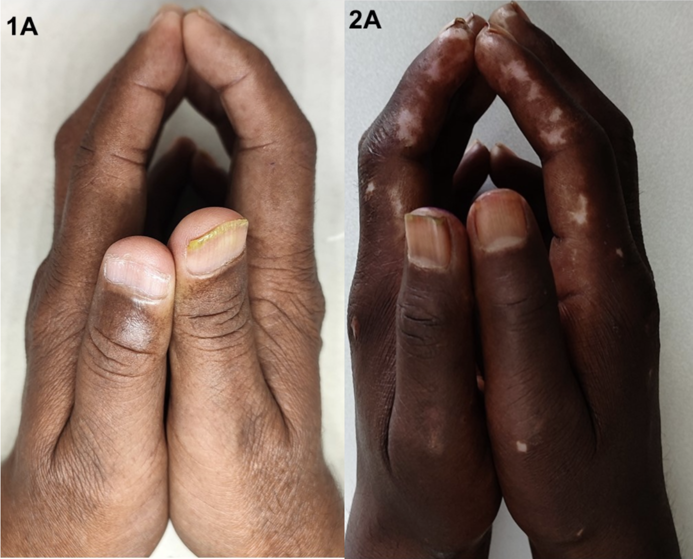
Limited Joint Mobility (LJM), or diabetic cheirarthropathy, is associated with both type 1 and type 2 diabetes mellitus. LJM presents as the “prayer sign”—inability to fully oppose the palms due to painless flexion contractures.1 Imaging typically reveals thickening of the palmar skin and flexor tendon sheaths without joint involvement.2,3 We juxtapose two patients with near-identical hand restriction: a 51-year-old man with type 2 diabetes and a 36-year-old woman with diffuse cutaneous systemic sclerosis (SSc). Both had marked limitation in finger extension (metacarpophalageal joint [MCP] extension limited to 5°) and a 40 mm gap between 5th MCPs. Hand function was better preserved in the patient with diabetes (Duruoz index 14 vs. 36). Axial proton density fat-saturated (PDFS) magnetic resonance imaging (MRI) at the carpal tunnel showed skin thickening, subcutaneous fat loss, and flexor tenosynovial thickening in both. In SSc,
more exuberant flexor proliferation (broad arrows), additional extensor tenosynovitis (narrow arrows), and more marked skin thickening were noted. Despite similar clinical findings, the MRIs reveal distinct differences. SSc shows clear inflammation, suggesting a dysregulated immune response driving fibrosis. In contrast, diabetes shows minimal inflammation, and the cause of hand stiffness is likely due to glucose-mediated collagen crosslinking. In SSc, skin fibrosis extent correlates with internal organ involvement and informs disease classification.4 It is tempting to speculate that even in diabetes, similar hand findings may signal a systemic fibrotic state.5 The humble “prayer sign” may thus serve as a visible clinical biomarker of fibrosis, offering a window into underlying systemic disease in both diabetes and SSc. Figure 1 (A-B). Prayer sign in a patient with type 2 diabetes mellitus; and Figure 2 (A-B) a patient with systemic sclerosis. MRI images show Axial proton density fat-saturated (PDFS) magnetic resonance imaging (MRI) of patient with contrast in tenosynovial thickening on the flexor aspect (broad arrows) and extensor aspect (narrow arrows).
Ethics declaration These participants are part of a study that has received ethical approval from the KEM Hospital Research Centre Ethics Committee (KEMHRC/RVC/EC/1518). Both patients have signed separate informed consent forms for photographs and MRI. Conflicts of interest The authors declare having no potential conflicts of interest.